Description
DESCRIPTION
Now used worldwide as an effective tool for intraoperative monitoring (IOM) of the spinal cord, the D185 MultiPulse Cortical Electrical Stimulator is the ONLY standalone surgical stimulator with FDA clearance for this technique.
The D185 MultiPulse Cortical Electrical Stimulator allows transcranial motor evoked potentials (MEPs) to be used in surgical procedures such as scoliosis correction, spinal tumour resection and thoraco-abdominal aortic aneurysm (TAAA) repair. The 1000V power source means that MEPs can even be evoked in patients with pre-existing neuropathologies.
The D185 MultiPulse Cortical Electrical Stimulator is also useful for peripheral nerve stimulation. Although the D185 was designed for transcranial cortical stimulation during intraoperative monitoring, the brief high voltage output also makes it suitable for use as a spinal root stimulator during differential diagnosis of peripheral nerve disorders, such as multifocal motor neuropathy and motor neuron disease. The high voltage allows effectively stimulation of deep nerve roots as they exit the spinal column, while the very short pulse duration minimizes patient discomfort.
Who Needs Intra-operative monitoring?
Surgical procedures carried out within or near the spinal column or those involving transient interruption of blood flow to the spinal cord (e.g repair of thoraco-abdominal aortic aneurysms) are associated with a risk of neurological impairment ranging from loss of sensation to complete paraplegia. These deficits can arise as a result of direct trauma, stretching of nerves or occlusion of blood flow. Much effort has therefore been made to develop techniques which allow the health of the spinal cord to be assessed continuously during these risky surgical procedures.
Sensory Evoked Potential (SEP) Monitoring
Surgical teams currently monitor the status of ascending spinal sensory pathways by applying stimuli to the patient’s ankle or wrist and observing the resultant changes in somatosensory evoked potentials (SEP’s) recorded from the brain. This form of intra-operative monitoring uses changes in the SEP waveform to alert medical teams of possible complications and there is no doubt that this technique has protected many patients from surgically induced neurological deficits. However, the technique of SEP monitoring has attracted some criticism, much of which has been published in peer-reviewed journals:
Unchanged SEP waveforms have on occasions misled surgeons into continuing with surgery, resulting in unforeseen post-operative neurological complications such as severe paraplegia.
Altered SEP’s have prompted surgeons to back-off from procedures, only to find that the patient has suffered no loss in sensory status upon recovery.
As SEP’s are generally small in magnitude, they can be difficult to monitor reliably in some patients, particularly those presenting with a pre-existing neuropathology.
Although SEP monitoring is used as an indicator of the health of the spinal cord as a whole, some would argue that for anatomical reasons, the descending motor fibres may be at greater risk during surgery. This would suggest that it would be of tremendous benefit to monitor descending motor fibres exclusively or in combination with SEP monitoring.
Transcranial Electrical Motor Evoked Potentials (tceMEPs) & Digitimer Ltd
In collaboration with leading clinical neurophysiologists, Digitimer developed the D185 MultiPulse Stimulator in order to provide a more reliable method of minimizing the risk of surgically induced paraplegia while maximizing the level of surgical correction that could be safely conducted. This unique device is used transcranially to electrically stimulate the brain’s motor cortex, resulting in a descending motor evoked potential (MEP) which is conducted down the spinal cord to upper and lower limb extremities. The pathways stimulated in this manner are the same as those used by the brain to trigger and control voluntary movement. As with SEP monitoring, any alterations in the MEP waveforms can provide the surgical staff with a crucial warning of possible complications.
MEP Monitoring – The Way Forward?
A 1000 patient, 2 centre clinical trial of the Digitimer D185 in the USA, demonstrated that MEP monitoring during spinal surgery was (1) more accurate for predicting motor outcome than the SEP was for predicting sensory outcome; and (2) that useful motor responses were achievable with a higher probability than useful sensory responses. Furthermore, in cases where SEP monitoring alone may have misled the surgeon into aborting or curtailing a procedure, additional use of MEP monitoring was shown to more reliably indicate whether it was safe for the surgeon to complete a procedure. Evidence from the 5-year study outlined above prompted the FDA to clear the Digitimer D185 MultiPulse Stimulator for marketing and the technique of MEP monitoring is now utilised extensively by cardiovascular and neurosurgeons all over the world.


DOWNLOADS
Product Information
MultiPulse Stimulator D185 Mk.IIa
D185 Electrode Holders
D185 MEP Connection Headboxes
Insertion of D185 Stimulus Output Connector
References
Publications which cite use of the Digitimer D185 can be found on Google Scholar.
DOWNLOAD BROCHURE

ACCESSORIES
Supplied
- Mains (Power) lead
- Operator’s Manual
Recommended
The D185 MultiPulse Cortical Electrical Stimulator can be supplied with a range of accessories to facilitate integration with your current operating theatre equipment and stimulation preferences. The current range includes electrode extension leads, stimulator output plugs, a footswitch and a range of electrode connection head boxes and electrode holders/handles for peripheral nerve stimulation applications.
Electrode Connection Headboxes
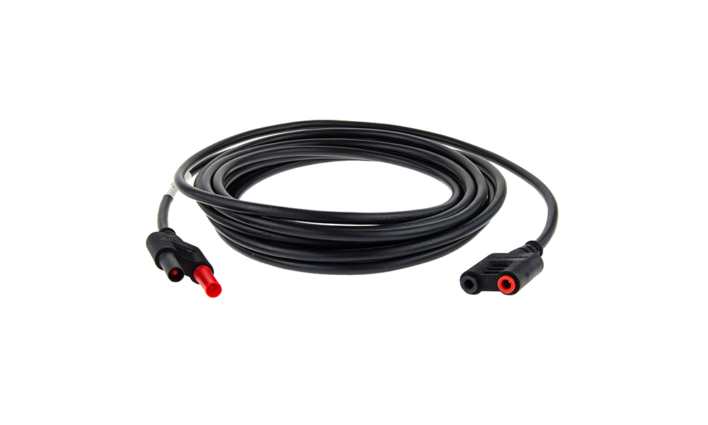
D185-HB4 Output Extension Cable
FEATURES
- Output extension cable for our clinical/human use stimulators
- Moulded connnectors at each end
- Robust cable of various lengths
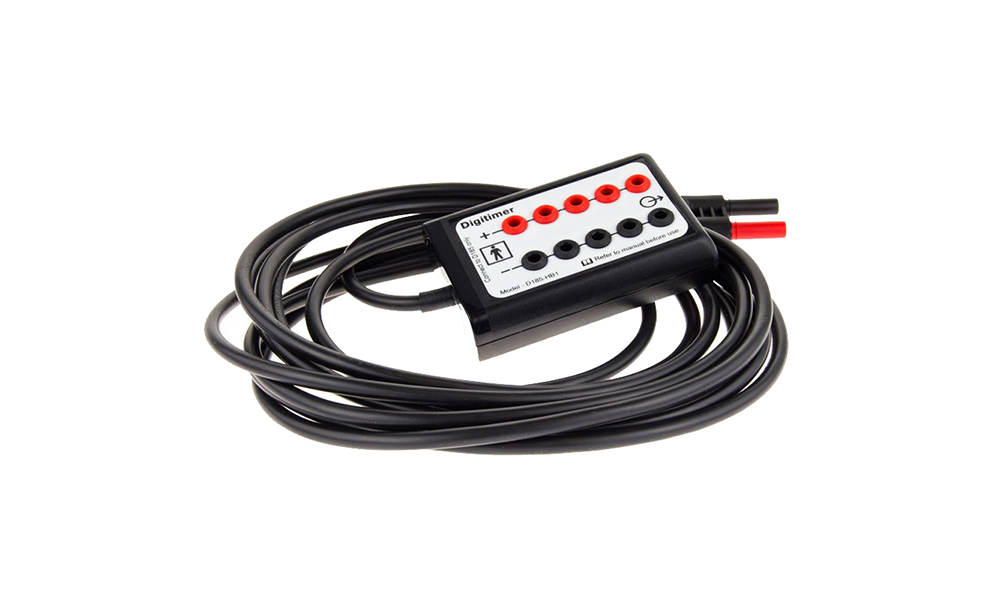
D185-HB1 Electrode Connection Headbox
DESCRIPTION D185-HB1 Electrode Connection Headbox (5m cable). This extension headbox provides 5 linked pairs of 1.5mm DIN sockets for connection
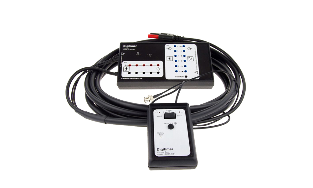
D185-HB3 Electrode Connection Headbox
D185-HB3 Electrode Connection Headbox with stimulus reversal and SEP electrode disconnection (inc. D185-CB1) – 5m. Ideal for those using SEP
Trigger Cables
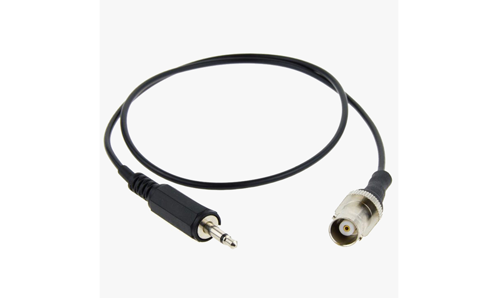
D185-TC5 Trigger Cable
D185-TC5 Trigger Cable (BNC socket – 3.5mm) – 0.6m. For connection between your EP system and the D185.
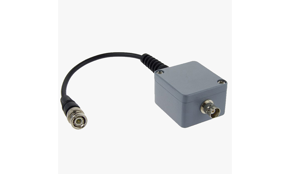
D185-TC4 Trigger Polarity Inverter
D185-TC4 Trigger Polarity Inverter. For connection between your EP system and the D185.
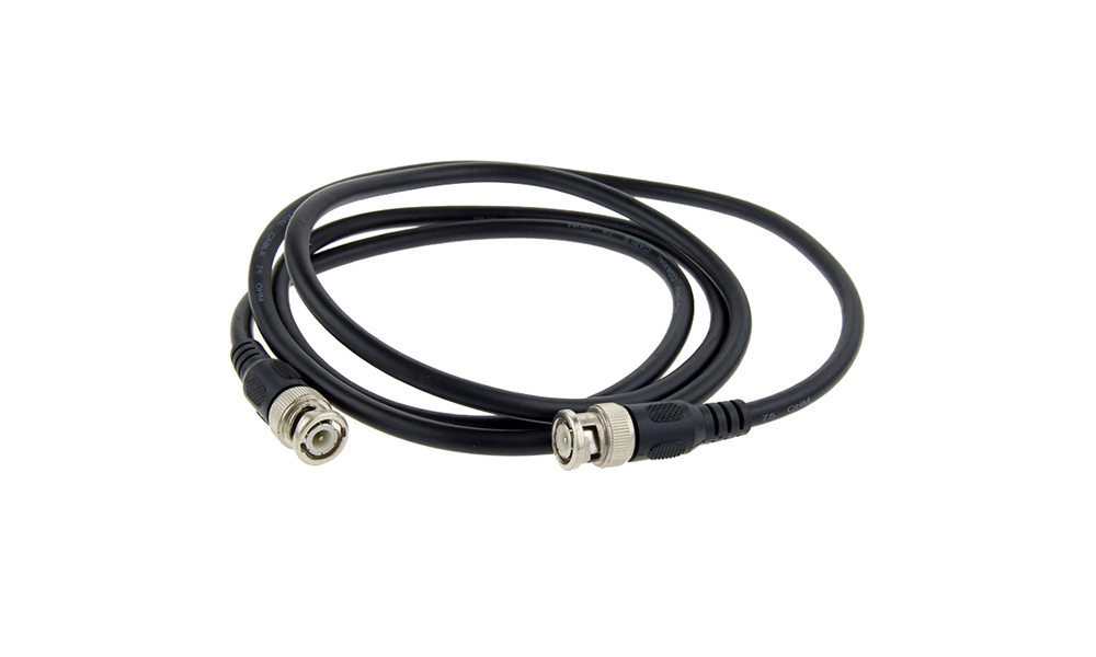
D185-TC3 BNC Trigger Cable
FEATURES
- Standard BNC based trigger cables
- Available in 1m or 2m lengths
- Suitable for multiple applications
- Compatible with our range of amplifiers and stimulators
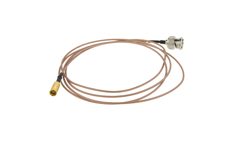
D185-TC2 Trigger Cable
D185-TC2 Trigger Cable (BNC – SMB) – 1.5m. For connection between your EP system and the D185.
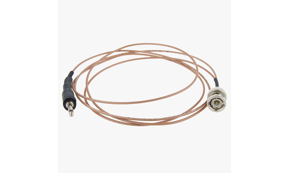
D185-TC1 Trigger Cable
DESCRIPTION D185-TC1 Trigger Cable (BNC – 3.5mm) – 1.5m. For connection between your EP system and the D185. GALLERY DOWNLOADS
Miscellaneous Items

D180-PADS Replacement Felt Pads
D180-Pads – Pack of ten 12mm felt pads for use with the D180 and D185 Electrode Handles.
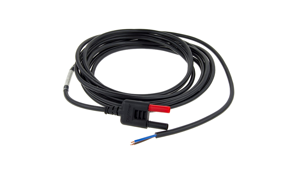
D185-OL1 Output Lead
D185-OL1 Output Lead – moulded connector on 5m cable for user assembly. Also suitable for use with the DS7A peripheral
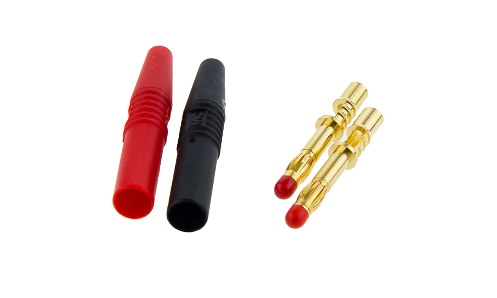
D185-OC1 Output Connector Plugs
D185-OC1 Output Connector Plugs for user assembly (pair). Also suitable for use with the DS7A peripheral nerve/muscle stimulator.
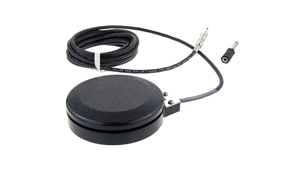
D185-FS1 Foot Switch
FEATURES
- Medical grade & sealed to IP68
- Supplied with 3.5mm to 1/4″ mono adaptor
- For use with D185, DS7 series and DS8R
Electrode Holders/Handles
D185-EH4 D180ES Style Electrode
D185-EH4 D180ES Style Electrode – Single anode and single cathode with long handle (with 60mm spacing).
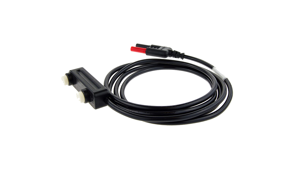
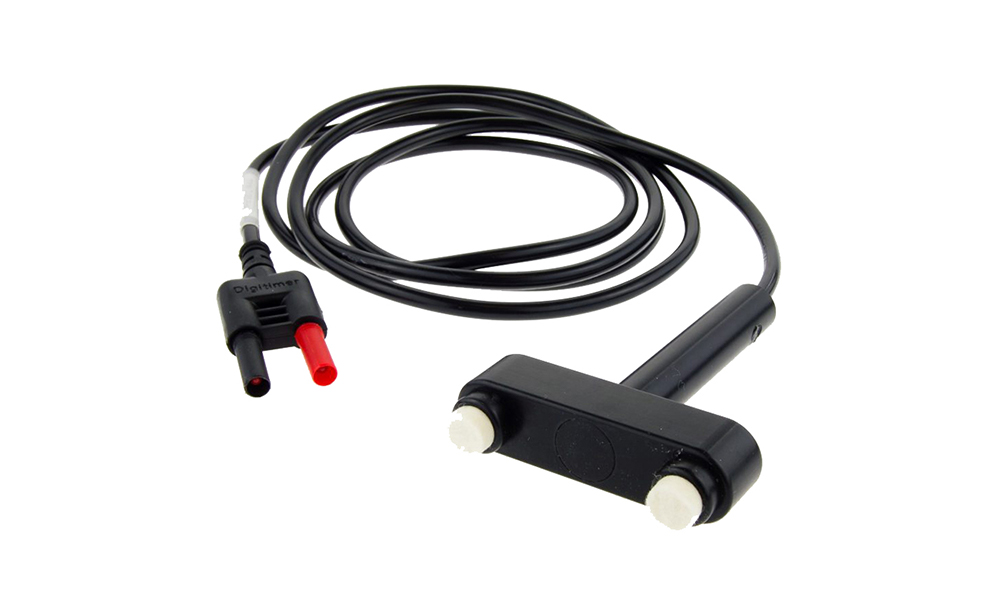
D185-EH3 Compact Standard Electrode
The D185-EH3 “Standard Electrode” is a small handheld electrode holder which features a single anode and cathode of 12mm diameter,
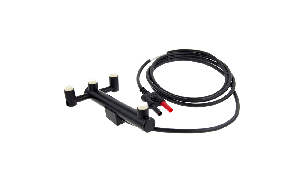
D185-EH2 Depth Electrode
D185-EH2 Depth Electrode – One cathode and three anodes for deep peripheral nerve stimulation.
FAQS
The D185 has certain safety limits that prevent excessive energy from being delivered to the patient. If you set the D185 to Normal mode and try to deliver too many pulses at too high a voltage, the unit will give an ERROR. You should consult the section titled Operating Modes in the D185 Operator’s Manual where you will find a graph that illustrated what these limits are.
Before you suspect any faults with the stimulator or cable, you should confirm that you have inserted the moulded plug of the extension cable correctly into the output socket on the front of the stimulator. When the plug mates with the output socket, it can be partly inserted with very little force, however, this does not provide a good electrical connection. For a complete electrical connection, further force is required and this supplement to the D185 users manual illustrates how the plug should be correctly inserted.
Yes, the D185-HB3 was specifically designed with this problem in mind. The D185-HB3 headbox incorporates 5 pairs of MEP output sockets as well as 6 channels of isolation for SEP electrodes. If the SEP electrodes are connected through the D185-HB3, they are briefly isolated while an MEP stimulus is passed. This prevents the EP system from “seeing” the MEP stimulus artefact and thus stops the amplifier from saturating/blocking for any length of time. You can read more about the D185-HB3 on the D185 page.
This is very much a decision that only you as the user can make, however we do supply a range of different accessories which start with the basic D185-HB4 which is essentially an output extension lead and end with the D185-HB3 which has 5 pairs of output sockets, a means of isolating SEP recording electrodes during MEP stimulus and a handheld trigger/polarity switching unit (D185-CB1) which allows the operator to easily trigger the D185. More detailed information on the accessories is available on the main D185 page.