Description
DESCRIPTION
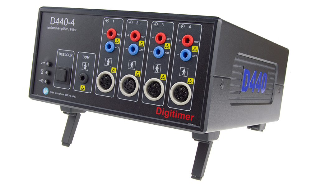
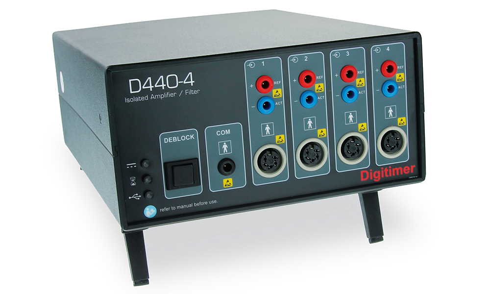
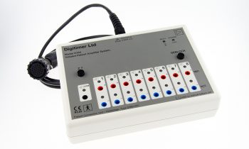
The Digitimer D440 Isolated EMG Amplifier is a a portable and standalone, low noise solution for human EMG studies, specifically those related to nerve excitability. The D440 features an amplification range of x100 to x20k. The gain, filter and mode settings for individual channels are adjusted using our own “virtual front panel” software or other software via a COM interface. The D440 is available in two versions, the D440-2 – a 2 Channel Isolated Amplifier – and the D440-4 – a 4 Channel Isolated Amplifier.
AC and DC Operating Modes
The D440 is designed to operate in AC and DC differential modes and includes a manually activated or externally gated de-block function, which can be useful for minimising the effects of magnetic stimulation artifacts.
Compatible with Standard Electrode Connectors
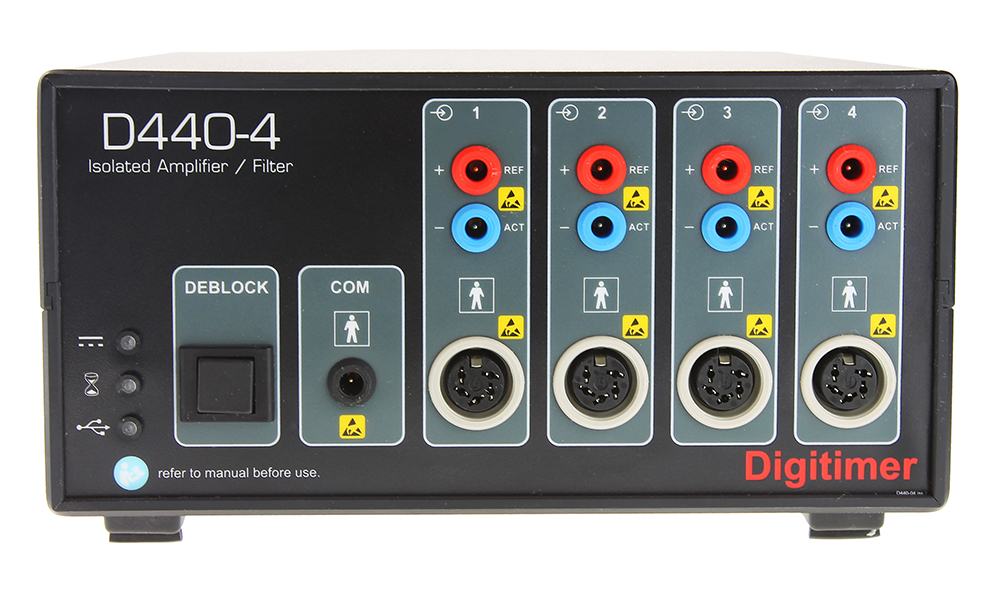
Electrodes are connected to the front panel via 1.5mm DIN 42 802 or standard 5-pole DIN connectors.
Designed for Human Research Applications
The D440 has been designed to meet international medical device design standards, however, it is NOT a medical device and its use is limited to human research studies only.
QtracW – Threshold Tracking & Nerve Excitability
The D440 has been designed to appeal to users of our DS5 Bipolar Constant Current Stimulator, who employ the DS5 and QtracW software to research human nerve excitability. This application requires a very low noise amplifier, which outputs an analog signal and can be controlled directly by the QtracW data acquisition software. These requirements are fully satisfied by the Digitimer D440 Isolated Amplifier.
Analogue Outputs for DAQ Hardware Compatibility
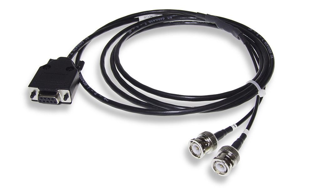
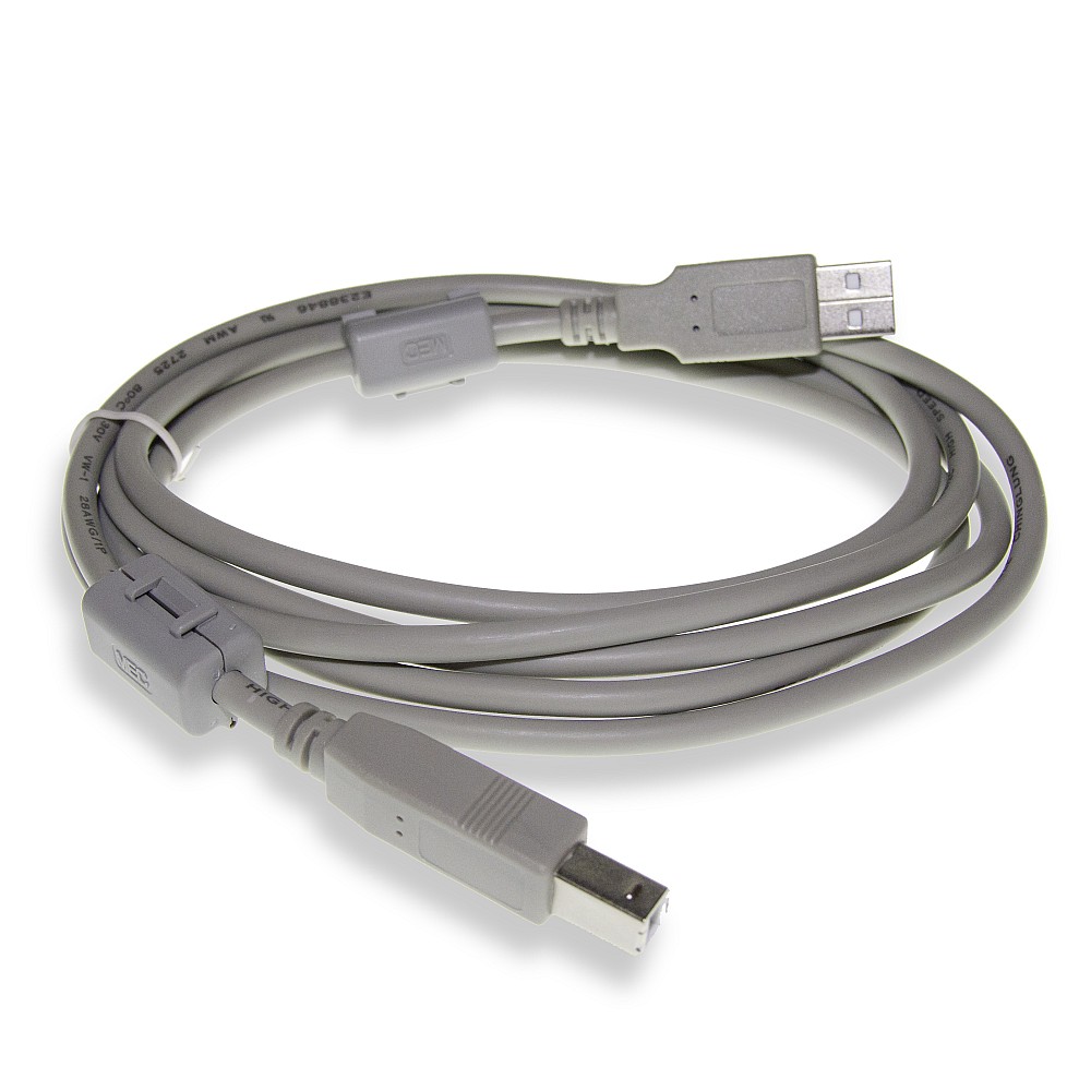
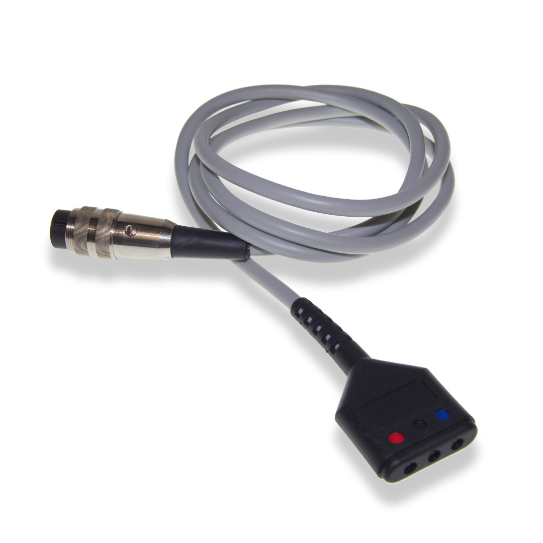
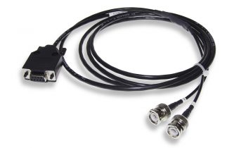
Each D440 amplifier is supplied with a signal output cable (D440-OL-xx, D connector to multiple BNC), electrode connection cable (D-440-IL, 1.2m long with 3x 1.5mm DIN42802 sockets for electrode connection and 270 degree 5-pin DIN plug for amplifier connection) and USB cable for connection to the host computer.
Features Overview
- Two (D440-2) or Four (D440-4) Channels of amplification, filtering and isolation – with independent control of each channel.
- Primarily designed as an AC amplifier, the D440 will also operate in DC mode.
- Input impedance of each channel is 1Gohm.
- On/off control of individual channels. The electronic inputs of individual channels can be grounded reducing cross-talk noise when recording from fewer channels. This also disconnects the subject from the electronics.
- Overall system GAIN for each channel x100 (10mV/V) to x20,000 (50μV/V).
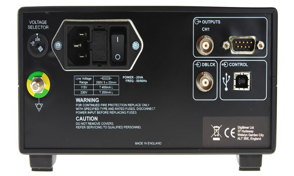
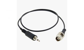
- Outputs have a ±5V range. The rear panel has a BNC socket for monitoring the output of channel 1 (this signal is mirrored on a 9-way ‘D’ connector on the rear panel along with the output signals of channels 2, 3 and 4). A signal output cable terminated with an appropriate number of BNC connections is supplied with each amplifier.
- LOW-CUT FILTER settings are selectable between 0 (DC), 0.159 Hz, 1Hz, 3Hz, 5Hz, 10Hz, 30 Hz and 50Hz for -3dB and are first order.
- HIGH-CUT FILTER settings are selectable between 1kHz, 3kHz, 5kHz, and 10kHz for -3dB and are second order, low phase shift Bessel style filters.
- The front panel contains three LEDs which are used to indicate the units power supply status, Internal-Error and Data-Bus Busy.
- The rear panel contains a mains IEC inlet socket with mains voltage selection, fuses and mains on/off switch, as well a 9-way “D” connector, for connecting channels to a data acquisition system and a USB port for connection to a Windows PC.
- A push button Deblock control is present on the front panel with a TTL compatible Deblock facility available via a BNC connector on the rear panel.
- Standalone Use – Previous amplifier settings will be maintained if a D440 is used without PC connection.
- Supplied with “virtual front panel” control software to adjust the settings of a single D440 amplifier. For applications requiring more than 4 channels, we recommend our D360 8-Channel Isolated Patient Amplifier.
- The D440 includes a COM interface to allow other software applications to control the amplifier settings.
- Mains operating voltage between 115V and 230V (switch selectable) at 50-60Hz. Note: for locations with low mains voltage (< 105V), such as some areas of Japan, a custom D440 with a replaced mains transformer will be required
GALLERY
PUBLICATIONS
Colomer-Poveda, D., Hortobágyi, T., Keller, M., Romero-Arenas, S., & Márquez, G. (2020). Training intensity-dependent increases in corticospinal but not intracortical excitability after acute strength training. Scandinavian Journal of Medicine and Science in Sports, 30(4), 652–661. https://doi.org/10.1111/sms.13608
Davies, J. L. (2020). Using transcranial magnetic stimulation to map the cortical representation of lower-limb muscles. Clinical Neurophysiology Practice. Elsevier. https://doi.org/10.1016/j.cnp.2020.04.001
Ghasemian-Shirvan, E., Farnad, L., Mosayebi-Samani, M., Verstraelen, S., Meesen, R. L. J., Kuo, M. F., & Nitsche, M. A. (2020). Age-related differences of motor cortex plasticity in adults: A transcranial direct current stimulation study. Brain Stimulation. Elsevier. https://doi.org/10.1016/j.brs.2020.09.004
Higashihara, M., Menon, P., van den Bos, M., Pavey, N., & Vucic, S. (2020). Reproducibility of motor unit number index and MScanFit motor unit number estimation across intrinsic hand muscles. Muscle and Nerve, 62(2), 192–200. https://doi.org/10.1002/mus.26839
Higashihara, M., Van den Bos, M. A. J., Menon, P., Kiernan, M. C., & Vucic, S. (2020). Interneuronal networks mediate cortical inhibition and facilitation. Clinical Neurophysiology, 131(5), 1000–1010. https://doi.org/10.1016/j.clinph.2020.02.012
Hossain, M. J., Kendig, M. D., Wild, B. M., Issar, T., Krishnan, A. V., Morris, M. J., & Arnold, R. (2020). Evidence of altered peripheral nerve function in a rodent model of diet-induced prediabetes. Biomedicines, 8(9). https://doi.org/10.3390/biomedicines8090313
Kiernan, M. C., Bostock, H., Park, S. B., Kaji, R., Krarup, C., Krishnan, A. V., … Burke, D. (2020). Measurement of axonal excitability: Consensus guidelines. Clinical Neurophysiology. Elsevier. https://doi.org/10.1016/j.clinph.2019.07.023
Mosayebi Samani, M., Agboada, D., Kuo, M. F., & Nitsche, M. A. (2020). Probing the relevance of repeated cathodal transcranial direct current stimulation over the primary motor cortex for prolongation of after-effects. Journal of Physiology. Wiley Online Library. https://doi.org/10.1113/JP278857
Mosayebi-Samani, M., Melo, L., Agboada, D., Nitsche, M. A., & Kuo, M. F. (2020). Ca2+ channel dynamics explain the nonlinear neuroplasticity induction by cathodal transcranial direct current stimulation over the primary motor cortex. European Neuropsychopharmacology, 38, 63–72. https://doi.org/10.1016/j.euroneuro.2020.07.011
Witt, A., Fuglsang-Frederiksen, A., Finnerup, N. B., Kasch, H., & Tankisi, H. (2020). Detecting peripheral motor nervous system involvement in chronic spinal cord injury using two novel methods: MScanFit MUNE and muscle velocity recovery cycles. Clinical Neurophysiology, 131(10), 2383–2392. https://doi.org/10.1016/j.clinph.2020.06.032
Witt, A., Bostock, H., Z’graggen, W. J., Tan, S. V., Kristensen, A. G., Kristensen, R. S., … Tankisi, H. (2020). Muscle velocity recovery cycles to examine muscle membrane properties. Journal of Visualized Experiments. jove.com. https://doi.org/10.3791/60788
Alaydin, H. C., Vuralli, D., Keceli, Y., Can, E., Cengiz, B., & Bolay, H. (2019). Reduced Short-Latency Afferent Inhibition Indicates Impaired Sensorimotor Integrity During Migraine Attacks. Headache, 59(6), 906–914. https://doi.org/10.1111/head.13554
Caetano, A., Pereira, M., & de Carvalho, M. (2019). A 15-minute session of direct current stimulation does not produce lasting changes in axonal excitability. Neurophysiologie Clinique, 49(4), 277–282. https://doi.org/10.1016/j.neucli.2019.05.067
Caetano, A., Pereira, P., Pereira, M., & de Carvalho, M. (2019). Modulation of sensory nerve fiber excitability by transcutaneous cathodal direct current stimulation. Neurophysiologie Clinique, 49(5), 385–390. https://doi.org/10.1016/j.neucli.2019.10.001
Colomer-Poveda, D., Romero-Arenas, S., Lundbye-Jensen, J., Hortobágyi, T., & Márquez, G. (2019). Contraction intensity-dependent variations in the responses to brain and corticospinal tract stimulation after a single session of resistance training in men. Journal of Applied Physiology, 127(4), 1128–1139. https://doi.org/10.1152/japplphysiol.01106.2018
Czesnik, D., Howells, J., Bartl, M., Veiz, E., Ketzler, R., Kemmet, O., … Paulus, W. (2019). I h contributes to increased motoneuron excitability in restless legs syndrome. Journal of Physiology, 597(2), 599–609. https://doi.org/10.1113/JP275341
Kristensen, A. G., Bostock, H., Finnerup, N. B., Andersen, H., Jensen, T. S., Gylfadottir, S., … Tankisi, H. (2019). Detection of early motor involvement in diabetic polyneuropathy using a novel MUNE method – MScanFit MUNE. Clinical Neurophysiology, 130(10), 1981–1987. https://doi.org/10.1016/j.clinph.2019.08.003
Kristensen, R. S., Bostock, H., Tan, S. V., Witt, A., Fuglsang-Frederiksen, A., Qerama, E., … Tankisi, H. (2019). MScanFit motor unit number estimation (MScan)and muscle velocity recovery cycle recordings in amyotrophic lateral sclerosis patients. Clinical Neurophysiology, 130(8), 1280–1288. https://doi.org/10.1016/j.clinph.2019.04.713
Mosayebi Samani, M., Agboada, D., Jamil, A., Kuo, M. F., & Nitsche, M. A. (2019). Titrating the neuroplastic effects of cathodal transcranial direct current stimulation (tDCS) over the primary motor cortex. Cortex, 119, 350–361. https://doi.org/10.1016/j.cortex.2019.04.016
Jacobsen, A. B., Kristensen, R. S., Witt, A., Kristensen, A. G., Duez, L., Beniczky, S., … Tankisi, H. (2018). The utility of motor unit number estimation methods versus quantitative motor unit potential analysis in diagnosis of ALS. Clinical Neurophysiology, 129(3), 646–653. https://doi.org/10.1016/j.clinph.2018.01.002
Jacobsen, A. B., Bostock, H., & Tankisi, H. (2018). Cmap scan mune (Mscan)-a novel motor unit number estimation (mune) method. Journal of Visualized Experiments. jove.com. https://doi.org/10.3791/56805
Makker, P. G. S., Matamala, J. M., Park, S. B., Lees, J. G., Kiernan, M. C., Burke, D., … Howells, J. (2018). A unified model of the excitability of mouse sensory and motor axons. Journal of the Peripheral Nervous System, 23(3), 159–173. https://doi.org/10.1111/jns.12278
Van Den Bos, M. A. J., Higashihara, M., Geevasinga, N., Menon, P., Kiernan, M. C., & Vucic, S. (2018). Imbalance of cortical facilitatory and inhibitory circuits underlies hyperexcitability in ALS. Neurology, 91(18), E1669–E1676. https://doi.org/10.1212/WNL.0000000000006438
Van den Bos, M. A. J., Menon, P., Howells, J., Geevasinga, N., Kiernan, M. C., & Vucic, S. (2018). Physiological processes underlying short interval intracortical facilitation in the human motor cortex. Frontiers in Neuroscience. frontiersin.org. https://doi.org/10.3389/fnins.2018.00240
Jacobsen, A. B., Bostock, H., Fuglsang-Frederiksen, A., Duez, L., Beniczky, S., Møller, A. T., … Tankisi, H. (2017). Reproducibility, and sensitivity to motor unit loss in amyotrophic lateral sclerosis, of a novel MUNE method: MScanFit MUNE. Clinical Neurophysiology, 128(7), 1380–1388. https://doi.org/10.1016/j.clinph.2017.03.045
ACCESSORIES
Supplied
- Mains Lead (Power Cord)
- Operator’s Manual
- Electrode Connection Input Lead (D440-IL, one per channel)
- Output Lead (D440-OL-2CH or D440-OL-4CH)
- USB Cable (D-USB-F)
- D440 Virtual Front Panel Control Software (supplied on USB Flash Drive)
