Description
DESCRIPTION
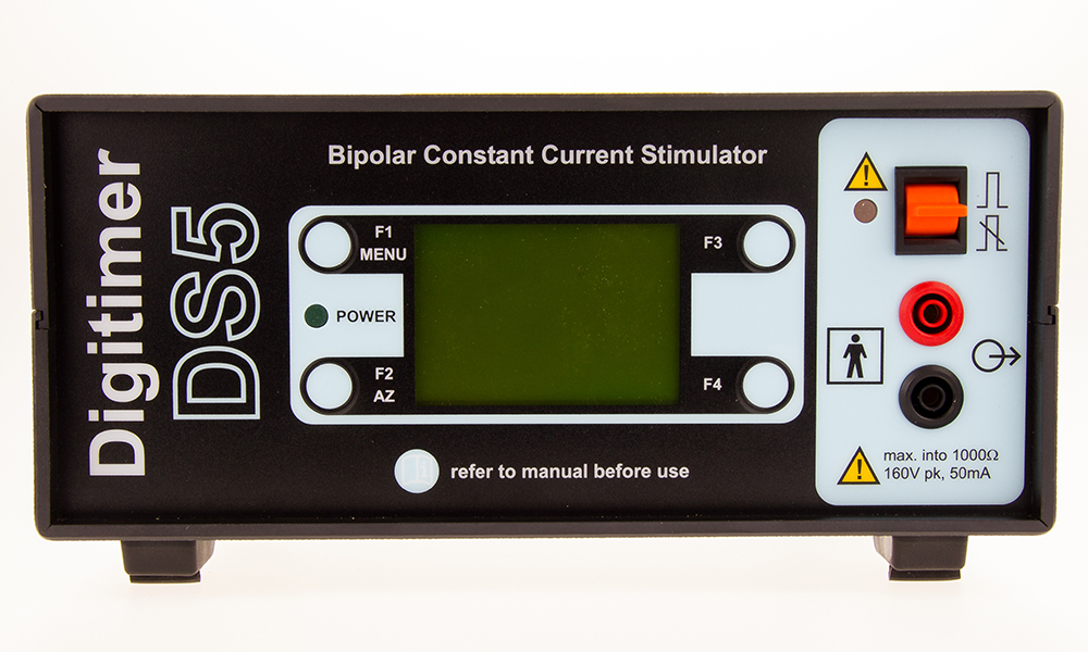
The DS5 Isolated Bipolar Constant Current Stimulator allows computer control of stimulus amplitude and timing parameters and has a maximum constant current output of ±50mA. It was originally designed to speed up and enhance human peripheral nerve diagnostics by facilitating semi-automated nerve excitability tests. However, it also has roles in wider aspects of clinical neurophysiology research, including psychological, vestibular system and nociceptive testing. More recently pairs of DS5’s have been used in research investigating the new technique of temporal interference stimulation (TIS), which may offer a non-invasive, but targeted alternative to deep brain stimulation and other therapies. The DS5 Isolated Bipolar Constant Current Stimulator is a CE marked medical device under the European Medical Device Regulation.
The DS5 is controlled by an analogue voltage input or “command” signal which it translates into an isolated constant current stimulus (up to ±50mA), precisely replicating the shape of the input waveform. As a result the DS5 should be of interest to anyone wishing to control surface electrical stimulation protocols via software/hardware combinations capable of producing a suitable command voltage waveform e.g semi-automated pain research or sensory threshold testing. The DS5 has also been employed for galvanic vestibular stimulation (GVS), transcranial AC stimulation (tACS) and temporal interference stimulation (tIS) protocols.
The DS5 was developed in collaboration with Prof. Hugh Bostock (UCL, London) for use with QtracW, a nerve excitability stimulus control, acquisition and data analysis software package, also available from Digitimer. This software semi-automates tests of nerve, muscle or cortical excitability using threshold tracking methods.
GALLERY
DOWNLOADS
DS5 Isolated Bipolar Stimulator Data Sheet
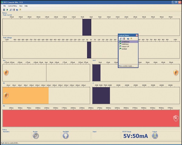
DS5 Control Software (Windows)
DS5 Control Software Installation Guide
PUBLICATIONS
The Digitimer DS5 Bipolar Constant Current Isolated Stimulator has been referenced in over 500 research papers, which can be viewed on Google Scholar. A few of the most highly cited papers published since 2019 are provided below.
Al, E., Iliopoulos, F., Forschack, N., Nierhaus, T., Grund, M., Motyka, P., … Villringer, A. (2020). Heart-brain interactions shape somatosensory perception and evoked potentials. Proceedings of the National Academy of Sciences of the United States of America, 117(19), 10575–10584. https://doi.org/10.1073/pnas.1915629117
Asamoah, B., Khatoun, A., & Mc Laughlin, M. (2019). tACS motor system effects can be caused by transcutaneous stimulation of peripheral nerves. Nature Communications. nature.com. https://doi.org/10.1038/s41467-018-08183-w
Engelmann, J. B., Meyer, F., Ruff, C. C., & Fehr, E. (2019). The neural circuitry of affect-induced distortions of trust. Science Advances. advances.sciencemag.org. https://doi.org/10.1126/sciadv.aau3413
Hird, E. J., Charalambous, C., El-Deredy, W., Jones, A. K., & Talmi, D. (2019). Boundary effects of expectation in human pain perception. Scientific Reports. nature.com. https://doi.org/10.1038/s41598-019-45811-x
Hoskin, R., Berzuini, C., Acosta-Kane, D., El-Deredy, W., Guo, H., & Talmi, D. (2019). Sensitivity to pain expectations: A Bayesian model of individual differences. Cognition. Elsevier. https://doi.org/10.1016/j.cognition.2018.08.022
Keywan, A., Jahn, K., & Wuehr, M. (2019). Noisy Galvanic Vestibular Stimulation Primarily Affects Otolith-Mediated Motion Perception. Neuroscience, 399, 161–166. https://doi.org/10.1016/j.neuroscience.2018.12.031
Sarigiannidis, I., Grillon, C., Ernst, M., Roiser, J. P., & Robinson, O. J. (2020). Anxiety makes time pass quicker while fear has no effect. Cognition. Elsevier. https://doi.org/10.1016/j.cognition.2019.104116
van Alst, T. M., Wachsmuth, L., Datunashvili, M., Albers, F., Just, N., Budde, T., & Faber, C. (2019). Anesthesia differentially modulates neuronal and vascular contributions to the BOLD signal. NeuroImage. Elsevier. https://doi.org/10.1016/j.neuroimage.2019.03.057
Wang, Y., Ge, J., Zhang, H., Wang, H., & Xie, X. (2020). Altruistic behaviors relieve physical pain. Proceedings of the National Academy of Sciences of the United States of America, 117(2), 950–958. https://doi.org/10.1073/pnas.1911861117
ACCESSORIES
Supplied
- Mains (Power) lead
- Operator’s Manual
- USB Cable
Recommended
- D185-HB4 Output Extension Cable
- D185-OC1 – Output Plugs
- Electrodes and electrode adaptor leads
FAQS
The DS5 has been designed to be as versatile as possible and therefore it should be compatible with most D/A hardware and software. The DS5 merely requires an analogue input of ±1, ±2.5, ±5 or ±10V at the BNC socket on the rear to provide the waveform which describes the stimulus. However, the user should note that they will need to source or write software that allows them to define the characteristics of the waveform.
Yes, we have developed GUI control software that allows this to be possible, you can download from this website.
No, after taking advice from collaborators we decided that a ±50mA limit was adequate for most cases, however, it is possible to link two DS5’s together in parallel to increase the overall stimulus output. Please contact us for details.
It is likely that the failure to re-enable the output is accompanied by a warning icon which indicates that re-enabling would result in DC stimulation. If this is the case, then the cause is almost certainly due to (i) a voltage waveform still being applied at the voltage input during the auto-zeroing procedure or (ii) noise being picked up through the input socket. For safety reasons, the DS5 will not re-enable if a significant stimulus current would immediately result.
This socket served two purposes – (i) It allows the operator to upgrade the firmware of the DS5 and (ii) it provides the communication link to control the DS5 settings from PC software. The DS5 User interface software can be downloaded from the Downloads tab on the DS5 product page. Do not connect the DS5 to the PC via the USB cable until this software has been installed.
The DS5 will identify the end point on a stimulation pulse as the stimulus current value falling to within ±400µA for a minimum period of 200µs or a sustained reversal of stimulus polarity (irrespective of the current amplitude). Therefore if you intend to carry out repetitive stimulation pulses of the same polarity, there needs to be a 200µs gap between them. If the pulses are of alternating polarity, there is no requirement for an interval (e.g as in a sine wave).
As with many electronic instruments, certain components within the DS5 make it important that the stimulator is switched ON and “warmed up” for at least an hour before it is used with a subject.
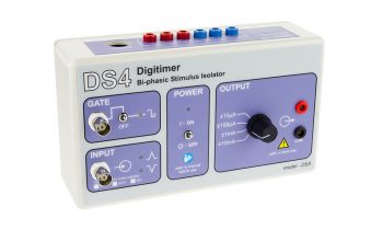
The DS5 is designed for use with human nerves in vivo and as such the sort of currents it generates are in the mA range. Even when the output of the DS5 has been recently autozeroed, some small currents (in the µA range) can persist and while these would be insignificant for human studies, they may be unwelcome in the rodent equivalent. Instead, we would recommend our DS4 stimulator for such non-human research studies.
The DS5 produces two types of audible alert. A high pitched “information” beep serves as confirmation that the operator has toggled or pressed a front panel switch or button. A deeper pitched “warning” tone informs the operator that a safety limit has been reached/exceeded or that intervention is required in response to a warning icon which will be simultaneously displayed on the front panel screen. Audible alerts are switched on by default, but the “information” beep may be disabled by the user via the options menu. When disabled a icon is displayed on the front panel LCD screen. If the DS5 beeps during stimulation, it is likely that there is a problem which the operator should investigate, such as an out of compliance error. The operator should check the LCD display for an information icon, which will explain why the warning is being given.
One of the requirements for our DS5 control software is the Microsoft VC++ 32bit, which is no longer included in new builds of Windows 10. As a result you will need to download and install it prior to installation of our DS5 software. If you cannot obtain it from Microsoft, we suggest you install the version of the DS5 software available here. This version includes Microsoft VC++ (32bit) within the installer.
The DS5 behaves like a voltage to current amplifier and is analogue throughout, so it does not have a resolution specification described in these terms. The main limitation to resolution is the noise level in the output signal, as that limits our ability to calibrate the DS5 as the current steps get smaller. However, we have data that confirms that the output of the DS5 can be effectively calibrated well below 100uA.

