NeuroLog System Application Note
NL100RK – Record, stimulate or lesion through one electrode

Overview
The Digitimer NeuroLog System is widely used to make extracellular field recordings from the brain and spinal cord, either in vivo or in slices. Once recordings are completed, it is often desirable to identify the exact recording site, so electrophysiological or pharmacological data can be aligned with anatomical knowledge. This may be accomplished by using electrical lesioning of the tissue, via the passage of an electrical current through the metal recording electrode. DC stimulation for several seconds leads to an electrolytic lesion that permits simple visualisation following staining. Also, when making gross nerve recordings, the ability to apply electrical stimulation through the recording electrode means that researchers can confirm the role or identity of the nerve being recorded from (Campbell et al. 2020).
Switch from recording to stimulation without risks of electrode movement
While it may be possible to disconnect the amplifier headstage in order to connect the voltage/current source used to make the lesion, this may be inconvenient or risk moving the electrode from the recording site. Instead, our NL100RK, combining the NL100RK Preamplifier Headstage & NL100C Stimulus Control Module, allows for combined recording/stimulation without having to disconnect the electrode from the amplifier. Simply replace our standard NL100AK headstage with the NL100AKS Headstage/NL100C Stimulus Control Module and add a stimulus source such as the Digitimer DS2A or DS3).
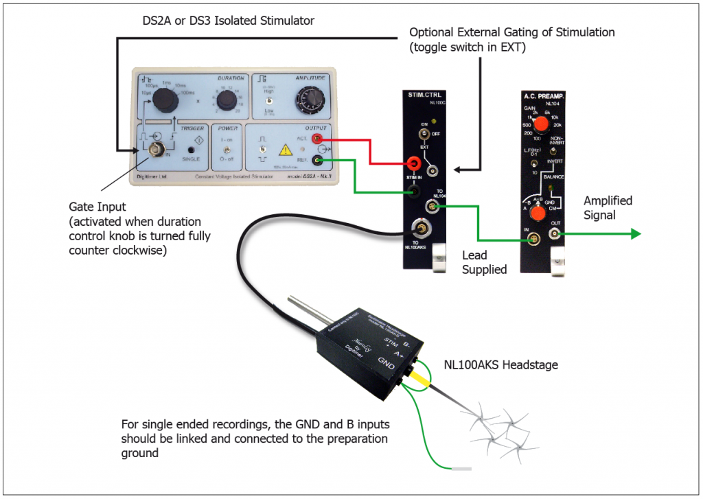
Use the NL100RK with stimulators including our DS2A and DS3
The NL100C will accept a voltage stimulus of up to 100V (e.g from our own DS2A stimulator shown above) and pass it through the NL100AKS headstage, subject to the position of the front panel toggle switch. In the “Off” position, stimuli are prevented from passing into the headstage and the preamplification circuitry operates, allowing an extracellular recording to be made. In the “On” position, any stimulus provided by the DS2A will be passed through the recording electrode to the preparation and the preamplification circuitry in the headstage is bypassed. In the “EXT” position, an external gating pulse can be used to control whether the headstage is in the stimulation or recording mode.
Further amplification and filtering of the amplified signal can be achieved by use of the NL106 AC/DC Amplifier and NL125/6 Filter. In addition, the NeuroLog System range includes the NL201 Spike Trigger and NL120S Audio Amplifier, which allow you to process your signal further and audibly monitor neuronal firing patterns.
Electrolytic lesioning of recording sites
The image above is an example of an electrolytic lesion in rodent spinal cord, provided by Dr Philip Holland (King’s College London). Dr Holland’s recordings were made using the NeuroLog System NL100RK and the electrolytic lesion was produced by the Digitimer DS3 Constant Current Isolator.
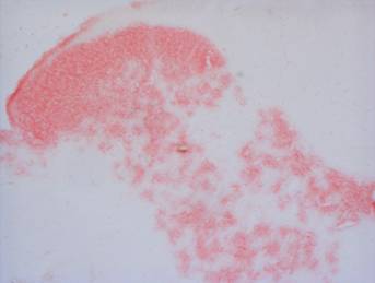
An internal jumper within the DS3 allows it to be set to deliver current while the “single-shot” trigger button was held. This allows for longer pulse durations than the front panel settings allow. Once electrophysiological recordings were completed, the spinal cord was stored in a solution of 10 % formaldehyde containing potassium hexacyanoferrate (III), which stains the iron deposits from the electrode black. The tissue was frozen and cut into 50 µm sections which were dry mounted on to gelatinised coated slides and counterstained with nuclear fast red to allow anatomical localisation of the lesion site.
To learn more about the modular Digitimer NeuroLog System or the NL100RK in particular, please check out the dedicated NeuroLog System section of the website or contact us.